

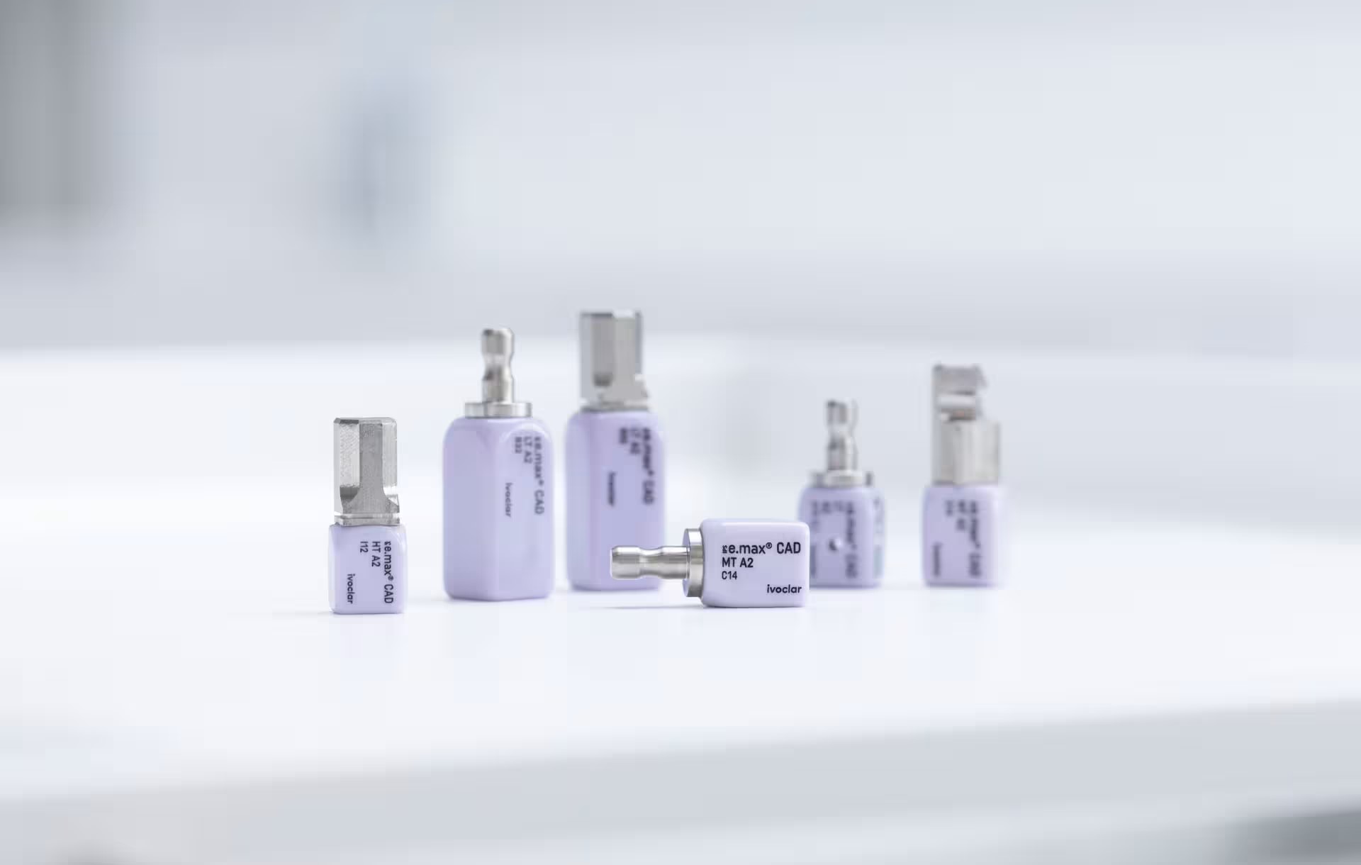
At the University of Washington, a 7-year prospective clinical investigation has provided valuable long-term insights into the performance of lithium disilicate CAD/CAM restorations in posterior teeth. Conducted by Prof. John A. Sorensen and his team, the study evaluated 50 IPS e.max CAD restorations fabricated with a fully digital workflow and adhesively bonded with Adhese Universal and Variolink Esthetic DC. With a Kaplan–Meier survival rate of 94.5% after up to seven years, the findings underline the reliability of adhesive, tooth conserving approaches in modern prosthodontics (Sorensen, Chen & Sadr 2025).
The advantages of lithium disilicate in posterior restorations.
Q&A with Dr. John Sorensen.
Q: From your clinical perspective, what are the key advantages of lithium disilicate ceramics such as IPS e.max CAD, especially for posterior restorations, compared to other ceramic material?
Sorensen: Above all, the conservation of tooth structure is most important. Adhesive ceramics like lithium disilicate allow us to preserve significantly more natural tooth structure than traditional full-crown restorations. In studies that Prof. Daniel Edelhoff and I conducted about 25 years ago, we demonstrated that even the most aggressive veneer design required just about half the tooth structure reduction of a full crown (Edelhoff & Sorensen 2002a). In posterior teeth, a 5-surface ceramic onlay design showed a similar result—roughly 50% less reduction (Edelhoff & Sorensen 2002b.).
We began with leucite-reinforced glass-ceramics, such as IPS Empress, which had a flexural strength of about 165 MPa and achieved excellent medium- and long-term results when adhesively cemented (Sorensen et al. 1998). With IPS e.max CAD lithium disilicate, we reached flexural strengths of around 475–500 MPa and improved fracture toughness. According to manufacturer data and confirmed by long-term internal quality testing over ten years, the average flexural strength of IPS e.max CAD is approximately 530 MPa. Adhesive cementation further enhances the final result by creating a ceramic/adhesive cement/tooth complex, conferring outstanding clinical longevity – even at reduced wall thickness.
Our in vitro and clinical data also show that properly polished lithium disilicate demonstrates wear behavior comparable to cast gold alloys (Sorensen et al. 2011; Sorensen 2013). And of course, its optical properties are unmatched – it simply looks like enamel. For single-tooth restorations, lithium disilicate remains the ideal choice.
Key factors for long-term adhesive success
Q: What are the most critical steps in adhesive cementation protocols to ensure the long-term success of conservative onlay preparations?
Sorensen: Before discussing bonding, we must ensure adequate ceramic thickness. For decades, my standard operating procedure has been to use the temporary restoration as the prototype for the final one. After occlusal adjustment, I measure thickness at key points using calipers to verify the preparation depth. In the case of milled restorations, we need to consider that the smallest milling bur is 0.6 mm – therefore, sharp internal line angles must be avoided to prevent overmilling.
When it comes to cementation, six factors are critical:
- Removal of temporary cement: Every trace must be removed; even residual pumice from cleaning can prevent complete seating.
- Isolation: Moisture control is non-negotiable. Saliva contamination after etching drastically reduces bond strength.
- Meticulous adhesive handling: Follow the manufacturer’s protocol exactly – etching time, active rubbing, air-thinning and curing. When using Adhese Universal, completely disperse the bonding agent with compressed air so no liquid remains, and be careful to avoid pooling in line angles.
- Avoid accumulation of excess adhesive: Since the protocol calls for photo-polymerization of adhesice prior to cementation, if excess material is cured it can prevent complete seating of the restoration
- Remove excess cement before curing: Since the 1990’s, even during our IPS empress and Empress 2 bridge studies (Sorensen et al. 1999), I have used micro brushes and waxed floss to remove excess before polymerization. If resin cement remains in the gingival embrasure, it can set and become nearly impossible to remove.
- Final Check: After curing, run unwaxed floss through contacts – if it frays that indicates that cement fragments remain and must be removed.
In my experience, these steps – though seemingly small – make the difference between average and excellent outcomes in adhesive restorations.
Clinical experience with Variolink Esthetic DC and Adhese Universal
Q: Based on your ling experience, how do you evaluate the long-term performance of Variolink Esthetic DC and Adhese Universal in highly stressed posterior areas?
Sorensen: My experience with Variolink cements spans mroe than 30 years. During my PhD studies in 1992-1997, I evaluated polymerization behavior in dual-curing and light-curing cements, and the original Variolink performed exceptionally well (Sorensen & Munksgaard 1995; 1996). Today, the modern Variolink Esthetic DC continues that legacy of reliability with improved esthetics and working stability. Even after prolonged storage, it maintains a consistent working time – essential when cementing multiple veneers or onlays simultaneously.
When assessing high-stress regions, such as second molars, which experience up to three times the bite force of central incisor, strength and adhesive bonding become decisive.
In our 7-year clinical study involving 35 onlays and 15 crowns, all fabricated digitally and cemented with Variolink Esthetic DC, we observed 94.5% clinical success rate, confirming the long-term reliability of this solution under posterior load. Incidentally, one of the two restoration occurrred by a patient biting into a stone in his food which fractured the onlay. The combination of IPS e.max CAD and proven adhesive cementation give clinicians confidence in their long-term outcomes.
Indications and clinical selection
Q: Which types of clinical cases are especially well-suited for adhesive cementation of lithium disilicate CAD?CAM restorations?
Sorensen: First, any tooth restoration that has not previously been treated with a full crown. My philosophy is simple: preserve enamel wherever possible. For anterior and premolar restorations, I typically use veneer-type preparations with 0.5-0.6 mm chamfer margins.
For posterior teeth with large composite fillings or cracks, especially mandibular molars with distal marginal ridge cracks (cracked tooth syndrome), lithium disilicate onlays are ideal. By restoring the structural integrity of posterior teeth adhesive ceramic onlays reinforce remaining tooth structure without aggressive preparation.
When blocking out undercuts, I use glass ionomer bases because their thermal expansioncoefficient is closest to dentin. It makes little sense to remove all internal geometry that improves retention and surface area for bonding.
Lithium disilicate is also the material of choice for full-mouth rehabilitiations requiring increased occlusal vertical dimension. Its high bond strength after hydroflouric acid etching, combined with enamel-like translucency, makese it ideal for conservative reconstructions.
Zirconia, even with MDP-based cements, typically achieves only 40-50% of the bond strength of etched lithium disilicate (Sorensen & Chen; Bitsis 2015).
Digital Workflow and marginal fidelity
Q: Your study also emphasized the advantages of a fully digital workflow. What are the most relevant findings in this regard?
Sorensen: The accuracy of modern digital workflow is remarkable. Using intraoral optical scanning (Trios 3, 3Shape), virtual design (Dental Design, 3Shape) and IPS e.max CAD milling, we produced restorations with mean marginal discrepancies of just 73 μm no supporting cusp shoulder margins and 53 μm on non-supporting cusp bevels, verified with optical coherence tomography (OCT) imaging (Sorensen, Hayashi & Sadr 2019). These are superior to many conventionally fabricated restorations.
In our up to 7-year trial, we also recorded time savings. In the middle third of cases, about 40% of restorations required no proximal or occlusal adjustment. In the final third, that number rose to 87% (Sorensen et al. 2025). Adjustments, when needed, typically took only one or two minutes.
This level of accuracy has huge implications for clinical efficiency. In early cases, we allocated an hour for delivery and cementation; later, most were finished in 15 minutes. These gains in productivity and predictability truly change the workflow for restorative dentistry.
As CAD/CAM systems evolve with faster milling and AI-assisted scanning, we can expect even greater efficiency and precision in the near future.
Future Trends
Q: What innovations or trends in adhesive protocols or CAD/CAM ceramics will have the greatest impact on restorative dentistry in the coming years?
Sorensen: The evolution of digital fixed prosthodontics continues at an impressive pace. Chairside systems are faster and more precise; software integration between clinicians and laboratories keeps improving.
The bigger paradigm shift, though, is the growing clinical confidence in conservative adhesive restorations. Many clinicians still choose full crowns, especially zirconia,
because it feels safer and simpler. Yet long-term data show that adhesively bonded ceramics, when used correctly, are equally reliable and far more tooth-conserving.
Several studies, including ours, indicate a significantly lower incidence of root canal therapy and postoperative sensitivity in adhesive onlays compared to full crowns
(Edelhoff & Sorensen 2002; Sorensen et al. 2022).
Our 7-year data clearly demonstrate that IPS e.max CAD restorations adhesively bonded with Adhese Universal and Variolink Esthetic DC provide stable adhesive interfaces, excellent marginal adaptation and reliable clinical performance even under posterior
stresses. I believe the combination of adhesive lithium disilicate ceramics and a precise digital workflow offers the perfect balance of strength, efficiency and the highest esthetics– and will remain a cornerstone of modern restorative practice for years to come.
Conclusion
The 7-year University of Washington study led by Prof. Sorensen offers robust clinical evidence for the long-term reliability of adhesive lithium disilicate restorations. With a 94.5% survival rate and minimal marginal degradation (Sorensen et al., IADR J Dent Res 2025 Ab #1214), IPS e.max CAD restorations cemented with Adhese Universal and Variolink Esthetic DC have demonstrated excellent durability, esthetics and bond stability – even in high-stress posterior applications.
As Prof. Sorensen emphasizes, the synergy of digital precision, adhesive strength and minimally invasive treatment approaches marks a new standard in fixed prosthodontics – one where efficiency and longevity go hand in hand with minimally invasive care.
We thank Prof. Sorensen for sharing his insights and long-term clinical experience in adhesive dentistry.
Image Overview

© John A. Sorensen. Posterior IPS e.max CAD onlay on tooth 14 (26) after 6.3 years in function
Posterior IPS e.max CAD onlay on tooth #14 after 6.3 years in function, demonstrating sustained surface gloss as well as stable stain and glaze characteristics.

© John A. Sorensen. Posterior IPS e.max CAD onlay on tooth 18 (37) after 7.2 years in function
Short clinical crown with limited retention and resistance form for full coverage. Restored with a conservative adhesively bonded ceramic onlay.


© John A. Sorensen. Posterior IPS e.max CAD onlay on tooth 30 (46) after 7.5 years in function (lingual and quadrant)

© Ivoclar IPS e.max CAD and Variolink Esthetic DC
References
Edelhoff D, Sorensen JA.Tooth structure removal associated with various preparation designs for anterior teeth. J Prosthet Dent 2002a;87:503–509.
Edelhoff D, Sorensen JA.Tooth structure removal associated with various preparation designs for posterior teeth. Int J Periodontics Restorative Dent 2002b;22:241–249.
Sorensen JA, Chen Y-W, Sadr A. Posterior CAD/CAM adhesive ceramic restorations: 7-year clinical study status. J Dent Res 2025; Abstract #1214
Sorensen JA, Hayashi J, Sadr A. Clinical trial of OCT imaging of posterior adhesive ceramic restorations. J Dent Res 2019;98:Abstract #2811.
Sorensen JA, Chen Y-W, Sadr A. Adhesive ceramics: 2-year clinical study results with a cracked tooth focus. J Dent Res 2022; Abstract #1007.
Sorensen JA, Choi C, Fanuscu MI, Kang SK, Mito WT. IPS Empress crown system: Threeyear clinical trial results. J Cal Dent Assoc 1998;26:130-136.
Sorensen JA, Cruz M, Mito WT, Raffeiner O, Meredith HR, Foser HP. A clinical investigation on three-unit fixed partial dentures fabricated with a lithium disilicate glassceramic.
Pract Periodont Aesthetic Dent 1999;11:95 -106
Sorensen JA, Munksgaard EC: Interfacial gaps of resin cemented ceramic inlays. Eur J Oral Sci 1995;103:116-120.
Sorensen JA, Munksgaard EC. Relative gap formation of resin-cemented ceramic inlays and dentin bonding agents. J Prosthet Dent 1996;76:374-378.
Sorensen JA, Chen Y-W. Shearbond strength of cements to dentin with varying moisture conditions. J Dent Res 2017;96:Spec.Iss.A, Ab#1616.
Sorensen JA, Sultan EA, Sorensen PN. Three-body wear of enamel against full crown ceramics. J Dent Res 2011;89 Ab#1652.
Sorensen JA. Finishing and polishing of modern ceramic systems. Inside Dentistry 2013;9(1) Special Issue 1:10-16
Bitsis I. Shear bond strength of adhesive cements to zirconia ceramics. MSD 2015, University of Washington.
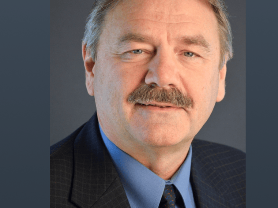
Dr. John A. Sorensen
Prof. John A. Sorensen, DMD, PhD, FACP, is Professor of Restorative Dentistry at the
University of Washington and Director of the B4T Laboratory. A diplomate of the
American Board of Prosthodontics, he focuses his research on adhesive dentistry,
CAD/CAM ceramics, and digital workflows in clinical prosthodontics.

You must be logged in to post a comment.