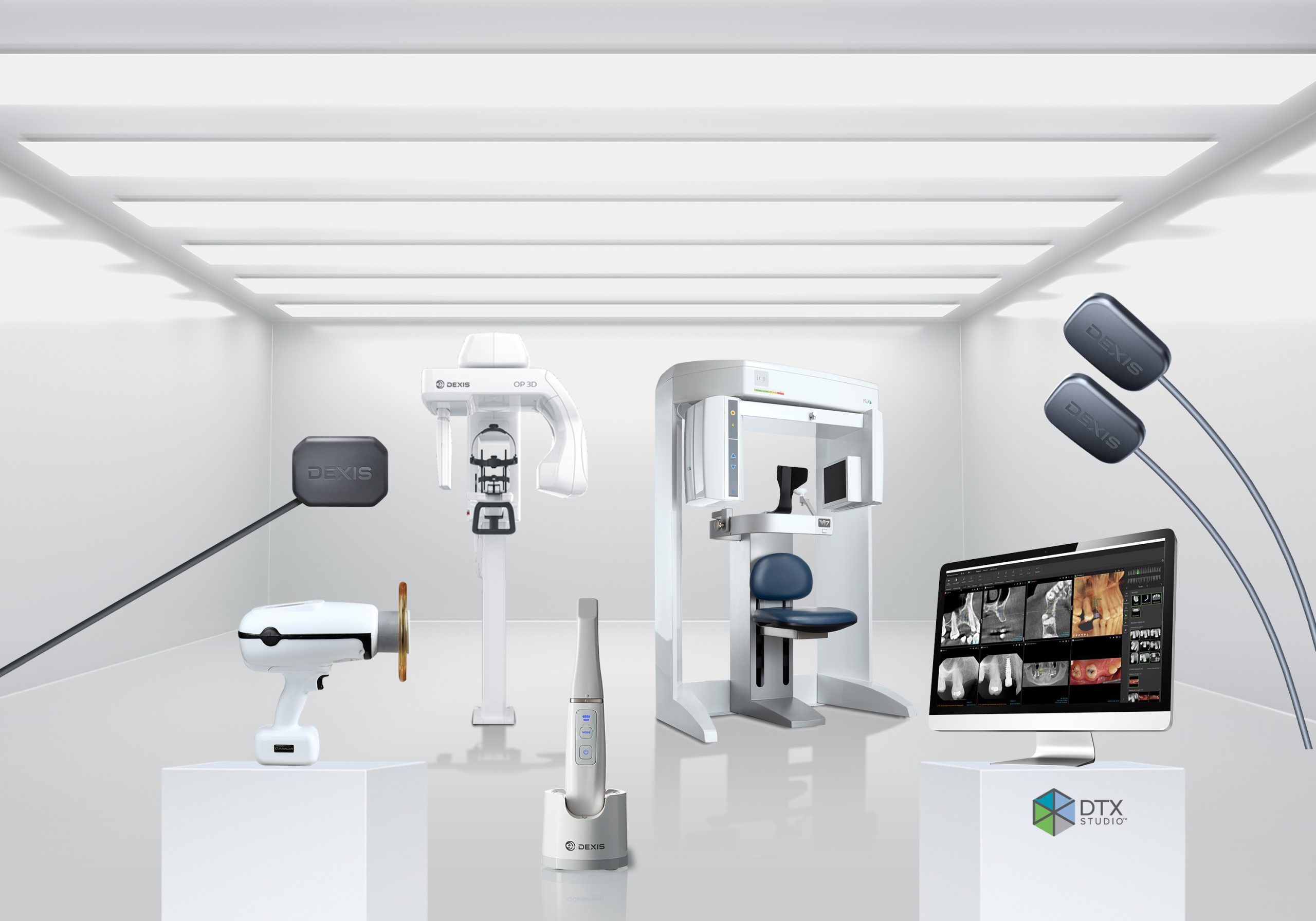
All content provided by DEXIS
The challenge: If I could see more, I could treat more. Effective treatment depends on detecting problems early. But poor image quality can delay diagnosis, leading to loss of additional tooth structure, endo treatment, retreatments, extractions, or failed implants. This can force you to remove more of the tooth structure when you do address the problem.
Image quality is only as good as the weakest part of your system. It can be impacted by many variables, including the sensor, software, and patient positioning. Using the wrong settings or a patient moving during the scan could also influence what you see.
Even if the quality is there, many clinicians struggle to bring multiple images together. You must devise a way to link disparate systems from multiple manufacturers into a single view. Once you import the files, you have to assemble them and often manually assign tooth numbers. And every minute you spend laying an intraoral scan over a CBCT image is time you aren’t at the chair with a patient. This all has a negative impact on patient care, your revenues, and ultimately the value of your practice.
But when you work with DEXIS products, you’ll be able to understand your patient better. Here’s how you’ll make it possible…
See in greater detail
With DEXIS products, you’ll generate consistent, high-quality images, using solutions that are comprehensively designed to help you discern the necessary pathology, including MAR, noise reduction and crisp filter options, improved bitewing view, and next generation adaptable pan scanning capabilities. In fact, our systems consistently win dentist choice awards, including 2022 Townie Choice Awards for the DEXIS Titanium intraoral sensor and 2022 Cellerant Best of Class Technology Award for DTX Studio™ Clinic software. What’s more, with our CBCT solutions, you’ll visualize all the internal structures noninvasively, enabling you to make an accurate diagnosis without opening the tooth. At the same time, with features such as our automatic exposure settings, you’ll make it easy for your staff to compensate for real-world conditions. And all of this is backed by the DEXIS 60-day satisfaction guarantee, along with ongoing training and education, so you can ensure your staff knows how to use your equipment properly.
This all means you’ll have the information needed to make the right diagnosis—and do so early— leading to less anxiety for you, your practice, and your patients.
 DEXIS imaging solutions are continually evolving with product updates and new DTX Studio Clinic releases.
Quickly assemble a complete view of the patient
You only have so many hours in the day. Minimize your manual steps and maximize your patient outcomes, with AI-powered DTX Studio Clinic software. Let DTX Studio Clinic’s MagicSort™ feature automatically recognize, sort, orient and organize your clinical photography as well as 2D X-rays, while you focus on your patient. Simplify mandibular nerve canal annotation with AI-enabled auto tracing, helping keep patients safe. Save time organizing your 3D case with Magic Detection auto setup of lower and upper panoramic curves, the TMJ workspace, Smart Focus, and even corrected patient orientation. Instantly import your intraoral scans with the tap of a button to complete your cases with single-screen views. Expand your anatomical views by AI-enabled merged CBCT and intraoral scans.
With one centralized location, you only need a single click to focus all of your X-rays, photos, 2D and 3D images, extraoral and intraoral images on a tooth (or region of interest). When AI-powered features control them all, you’ll have everything you need to capture the efficiencies of digital dentistry. And transform patient smiles.
Simplify your analysis
You want it to be as easy as possible to discover what’s going on with your patients. But supporting multiple systems that don’t talk to each other takes you away from this goal. With the DEXIS full suite of solutions, you’ll reduce the number of standalone applications you use, creating greater connectivity and patient image integration across your office. When you see more of the patient’s anatomy, you can be more confident in your diagnosis and treatment. At the same time, you’ll facilitate the sharing of images with third-party labs or specialist colleagues. You’ll also reduce the number of contact points when you have questions or require technical support. And with fewer systems to learn, you’ll minimize the associated downtime for you and your staff.
Most importantly, you can seamlessly combine all the images related to the patient into a single, coherent view—sensor, camera, panoramic, 3D, and intraoral scans—capturing all the information needed to feel more confident in your diagnosis.
“My cone beam and my DTX aren’t going anywhere. It’s the bedrock of my practice.”
Gary Williams, DMD, Williams Dental and Associates
Read his success story: https://www.pattersondental.com/resources/dental-equipment/digital-imaging-williams-dental
Learn more: https://www.pattersondental.com/equipment-technology/digital-imaging/extraoral/dexis-op-3d


You must be logged in to post a comment.